Photoacoustic (PA) imaging modalities are internationally regarded as a very promising complement or alternative to conventional magnetic resonance imaging (MRI), positron emission tomography (PET), X-rays, ultrasound and optical (“biophotonic”) imaging. During the past 7 years we developed [1-3] a patented [4] and patent-pending [5] “Photoacoustic Radar” (PAR) technology that does not require nanosecond pulses and bulky, expensive Q-switched lasers. We demonstrated that the PAR can be used with a standard ultrasonic (US) phased array for B-mode imaging similar to conventional ultrasound [6]. The similarity between conventional US and PAR signal generation and processing, as well as the superior subsurface localization of the chromophore exhibited by the PAR phase image, have no parallel in today’s widely used pulsed-laser PA technologies. We demonstrated US and PAR image co-registration of mouse tumors for easy identification of the location of the tumor (US) and morphological details of the spatial blood distribution in the tumor (PAR) [7].
We showed that the PAR can be used with a standard ultrasonic phased array for B-mode imaging similar to conventional ultrasound (US) and can image blood vessels in the wrist of a human volunteer [8,9].
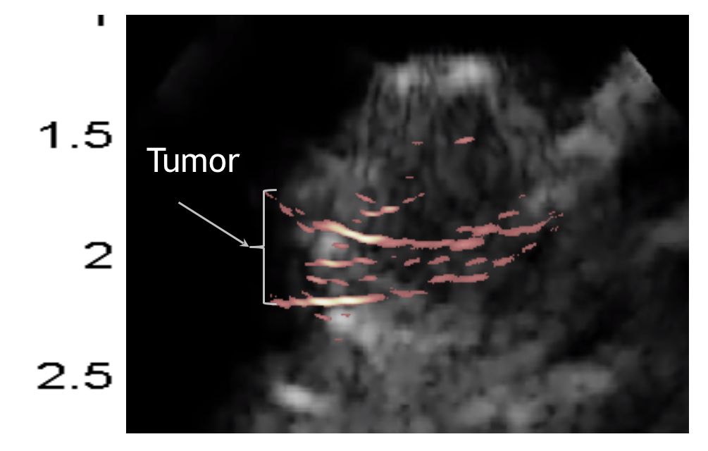
The figure above shows the superposition of US (background) and PAR (“Tumor”) phase images from a tumor grown inside the thigh of a mouse (Y-axis: depth, cm). Image co-registration yields easy identification of the tumor location (US) and morphological details of its spatial blood distribution in the tumor (PAR).
The figure above shows (a) a Phase-filtered PAR image superimposed on the pure US image of the left thigh of a mouse injected with a tumor grown inside the thigh of a mouse (Y-axis: depth, cm). (b) Zoomed image of the region of interest from Image co-registration yields easy identification of the tumor location (US) and morphological details of its spatial blood distribution in the tumor (PAR).
PAR impact: Biomedical photoacoustic radar imagers constitute paradigms of integration at the interface between imaging instrumentation, signal generation physics, and biophotonics. They have distinct attributes and the potential for rapid penetration into the growing pre-clinical small animal in-vivo testing field, and ultimately, in clinical settings for diagnosis of multiple diseases like cancer. They feature (1) high image acquisition frame rates (kHz) capable of practical real-time clinical imaging, (2) small inexpensive laser diode source footprints with the potential for portable multispectral imaging applications, (3) two images (amplitude and phase) at each probed subsurface depth for higher diagnostic power, (4) depth selectivity via cross-correlation delay-time fixing (quick tomographic slice localization and operator-controlled fixed-depth image formation), (5) mm-level axial resolution, and (6) potential for reliable diagnosis of pre-malignant tumors and early cancers based on quantitative images using wavelength-modulated differential schemes and phase filtering. Currently, the PAR is being developed for intravascular imaging of the coronary artery using a catheter in partnership with Colibri Technologies, Inc. (Toronto).
References
[1] Telenkov S, Mandelis A. Frequency-domain photothermoacoustics: alternative imaging modality of biological tissues. J Appl Phys 2009; 105: 102029.
[2] Telenkov S, Mandelis A. Photothermoacoustic imaging of biological tissues: maximum depth characterization comparison of time and frequency-domain measurements. J Biomed Opt 2009; 14: 044025.
[3] Telenkov S, Mandelis A. Signal-to-noise analysis of biomedical photoacoustic measurements in time and frequency domains. Rev Sci Instrum 2010; 81: 124901.
[4] Mandelis A, Vitkin A, Telenkov S, and Fan Y. (inventors); “Laser Photo-Thermo-Acoustic Imaging Frequency-Swept Heterodyne Lock-in Instrumentation for Industrial and Biomedical Materials”, US patent 7,525,661 B2, Issued: April 28, 2009.
[5] Mandelis A, Telenkov S, Lashkari B. (Inventors). Systems and Methods for Frequency-Domain Photoacoustic Radar Phased-Array Imaging. (US patent application 13/660,771, filed October 25, 2012).
[6] Telenkov S, Alwi R, Mandelis A, Worthington A. Frequency-domain photoacoustic phased array probe for biomedical imaging applications. Opt Lett 2011; 36 (23): 4560-2.
[7] Dovlo E, Lashkari B, Mandelis A, Shi W, Liu F-F, Photoacoustic Radar Phase-Filtered Spatial Resolution and Co-registered Ultrasound Image Enhancement for Tumor Detection, Biomed. Optics Express 2015; 6 (3) 1003 – 1009.
[8] Telenkov S, Alwi R, Mandelis A, Worthington A. Frequency-domain photoacoustic phased array probe for biomedical imaging applications. Opt Lett 2011; 36 (23): 4560-2.
[9] Mandelis A, Telenkov S, Lashkari B. (Inventors). Systems and Methods for Frequency-Domain Photoacoustic Radar Phased-Array Imaging. (US patent application 13/660,771, filed October 25, 2012).