Osteoporosis (OP) is a major public health problem in Canada and around the world. Fractures from osteoporosis are more common than heart attack, stroke and breast cancer combined [1]. In preliminary studies we demonstrated the feasibility of photoacoustic detection of bone density variations [2]. Correlations between PAR and US modalities with the mineral volume fraction of the bone tissue were found [3]. The results showed that both PAR and US raster scanned images are sensitive to mineral changes. Furthermore, the PA modality is also sensitive to changes in the collagen content of the bone, but US is not statistically sensitive to these changes [4]. Impact: Considering that the prevailing OP diagnostic methods such as dual X-ray absorptiometry (DEXA) are only body-mass-index (BMD) specific, combination of PAR and US probes may result in improved sensitivity and specificity for the detection of bone demineralization or decollagenization, leading to more accurate and safer (than ionizing DEXA) bone loss diagnosis. In recent years we founded the SPIE annual BiOS (Photonics West) conference “Optics in Bone Surgery and Diagnostics”.
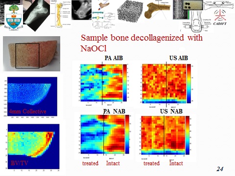
AIB: Apparent Integrated Backscattering/Back-propagating signal
NAB: Normalized Apparent Backscattering/Back-propagating signal
References I.3
[1] Osteoporosis Canada: www.osteoporosis.ca/osteoporosis-and-you/osteoporosis-facts-and-statistics/
[2] Lashkari B, Mandelis A. Co-registered photoacoustic and ultrasonic signatures of early bone density variations, J. Biomed. Opt. 2014; 19, 036015.
[3] Yang L, Lashkari B, Tan JWY, Mandelis A. Photoacoustic and ultrasound imaging of cancellous bone tissue , J. Biomed. Opt. 20(7), 076016 (12 pages) (2015). doi:10.1117/1.JBO.20.7.076016.
[4] Lashkari B, Yang L, Mandelis A, The Application of Backscattered Ultrasound and Photoacoustic Signals for Assessment of Bone Collagen and Mineral Content”, Quant. Imaging Med. Surg.; Special issue “Emerging Technologies in Biomedical Imaging”2015; 5(1), 46 – 56.