Pre-clinical imaging technologies are excellent tools for assessment of anatomical, physiological and functional parameters at molecular and cellular levels in live animal models. They play a significant role in the assessment of specific drug candidate or treatment regimes, and in phenotyping and understanding the pathophysiology of various diseases. Markets and Markets [1] reported that the overall pre-clinical in-vivo imaging market was valued at US $790 million in 2012 and estimated to grow at an annual rate of 14.5% until 2017. The report stated, however, that the high cost of imaging systems and technological limitations restrain market growth. Our recent development of Truncated-Correlation Photothermal Coherence Tomography (TC-PCT) presents a unique opportunity to introduce a new, relatively inexpensive, portable, three-dimensional, small-animal functional imaging technology. Two key functional imaging areas are being investigated: a) Hypoxia imaging in the blood of living mice/rats (brain vessel monitoring) will assess the sensitivity of the technique to tumor hypoxia, closely associated with tumor aggressiveness and therefore a key parameter to be monitored.
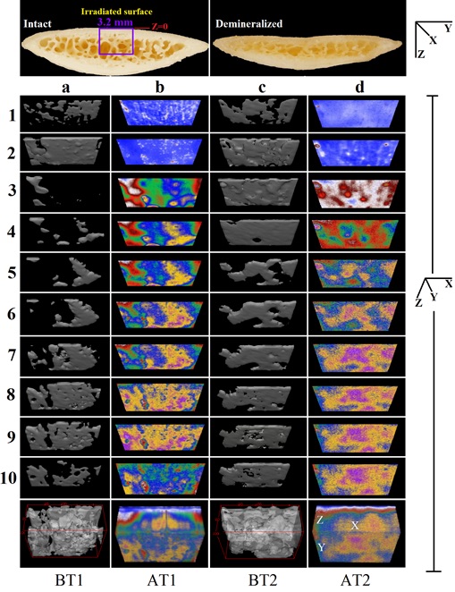
Binarized amplitude segments (a, c) and layer-by-layer planar TC-PCT images (b, d). The 10 binarized sequences, ~300 μm thick each, compose tomograms BT1 and BT2. The 120 planar sequences, ~25 μm thick each, constitute the absolute amplitude tomograms AT1 and AT2 [7]
Photoacoustic imaging of the brain is based on the acoustic detection of optical absorption from tissue chromophores, such as oxy-hemoglobin (HbO2) and deoxy-hemoglobin (Hb) [2]. It can simultaneously provide high-resolution images of mouse brain vasculature and hemodynamics with intact scalp [3]. b) Angiogenetic blood accumulation imaging in thighs of immunodeficient mice subjected to tumor injections. Two types of cancer cell lines (hypopharyngeal carcinoma FaDu and MDA-MB-231) will be used for relatively fast growth as in our recent PAR studies [4]. TC-PCT has the potential to complement or replace photoacoustic tomography (PAT) [5] with a more economical, compact, convenient and faster in-vivo mouse and rat imager with higher lateral and axial resolution and no raster scanning. TC-PCT Impact: The new Photothermal Coherence Imaging technology is expected to have substantial advantages over the fast growing PAT: no water interface (air coupled operation), no moving/rotating parts (time sequential slice images with stationary object), improved lateral resolution (100 μm vs. ~ 500 μm) and axial resolution (25 μm vs. 100 μm), and higher penetration depth in hard (boney) tissues (3.2 – 4 mm vs. 1.3 – 3 mm) [6].
References I.4
[1] http://www.marketsandmarkets.com/Market-Reports/pre-clinical-molecular-imaging-market-841.html
[2] Yang JM, et al., Nat Med. 2012; 18(8), 1297.
[3] Yao J, et al., Neuroimage 2013; 64, 257.
[4] Telenkov S, Alwi R, Mandelis A, Worthington A. Frequency-domain photoacoustic phased array probe for biomedical imaging applications. Opt Lett 2011; 36 (23): 4560-2.
[5] Gerling M, et al., Theranostics 2014; 4 (6), 604.
[6] Kaiplavil S, Mandelis A. Truncated-correlation photothermal coherence tomography for deep subsurface analysis, Nature Photonics 2014, 8, 635-642 / DOI: 10.10.1038/NPHOTON.2014.111.
[7] Kaiplavil S, Mandelis A, Amaechi BA. Truncated-correlation photothermal coherence tomography (TC-PCT) of artificially demineralized animal bones: 2- and 3-dimensional markers for mineral-loss monitoring, J. Biomed. Opt. 2014; 19(2), 026015.